Any other treatments will make the asymmetry worse!
2월 1, 2023
Asymmetric expansion
2월 1, 2023Changes in the midpalatal and pterygopalatine sutures induced by micro-implant-supported skeletal expander, analyzed with a novel 3D method based on CBCT imaging
Changes in the midpalatal and pterygopalatine sutures induced by micro-implant-supported skeletal expander, analyzed with a novel 3D method based on CBCT imaging
Background
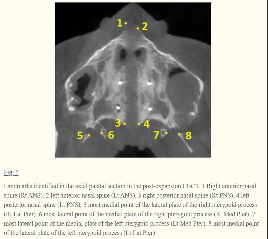
Mini-implant-assisted rapid palatal expansion (MARPE) appliances have been developed with the aim to enhance the orthopedic effect induced by rapid maxillary expansion (RME). Maxillary Skeletal Expander (MSE) is a particular type of MARPE appliance characterized by the presence of four mini-implants positioned in the posterior part of the palate with bi-cortical engagement. The aim of the present study is to evaluate the MSE effects on the midpalatal and pterygopalatine sutures in late adolescents, using high-resolution CBCT. Specific aims are to define the magnitude and sagittal parallelism of midpalatal suture opening, to measure the extent of transverse asymmetry of split, and to illustrate the possibility of splitting the pterygopalatine suture.
Conclusions
Midpalatal suture was successfully split by MSE in late adolescents, and the opening was almost perfectly parallel in a sagittal direction. Regarding the extent of transverse asymmetry of the split, on average one half of ANS moved more than the contralateral one by 1.1 mm. Pterygopalatine suture was split in its lower region by MSE, as the pyramidal process was pulled out from the pterygoid process. Patient gender and age had a negligible influence on suture opening for the age group considered in the study.
Many people have LEFT YAW on their maxilla. This is due to the LEFT YAW of the sphenoid bone. So the right ANS is a little more forward. This indicates that the maxilla is twisted and extended. To avoid this in all patients, the LEFT YAW of the sphenoid bone should be treated before expansion with MARPE. So, before MARPE, either cranial osteopathy or mcb splint treatment is required.
